New Insights Into Drug Discovery Targeting Tau Protein
Microtubule-associated protein tau is characterized by the fact that it is an intrinsically disordered protein due to its lack of a stable conformation and high flexibility. Intracellular inclusions of fibrillar forms of tau with a b-sheet structure accumulate in the brain of patients with Alzheimer’s disease and other tauopathies. Accordingly, detachment of tau from microtubules and transition of tau from a disordered state to an abnormally aggregated state are essential events preceding the onset of tau-related diseases. Many reports have shown that this transition is caused by post-translational modifications, including hyperphosphorylation and acetylation. The misfolded tau is self-assembled and forms a tau oligomer before the appearance of tau inclusions. Animal and pathological studies using human samples have demonstrated that tau oligomer formation contributes to neuronal loss. During the progression of tauopathies, tau seeds are released from cells and incorporated into other cells, leading to the propagation of pathological tau aggregation. Accumulating evidence suggests several potential approaches for blocking tau-mediated toxicity: (1) direct inhibition of pathological tau aggregation and (2) inhibition of tau post-translational modifications that occur prior to pathological tau aggregation, (3) inhibition of tau propagation and (4) stabilization of microtubules. In addition to traditional low-molecular-weight compounds, newer drug discovery approaches such as the development of medium-molecular-weight drugs (peptide- or oligonucleotide-based drugs) and high-molecular-weight drugs (antibody-based drugs) provide alternative pathways to preventing the formation of abnormal tau. Of particular interest are recent studies suggesting that tau droplet formation by liquid-liquid phase separation may be the initial step in aberrant tau aggregation, as well results that implicate roles for tau in dendritic and nuclear functions. Here, we review themechanisms through which drugs can target tau and consider recent clinical trials for the treatment of tauopathies. In addition, we discuss the utility of these newer strategies and propose future directions for research on tau-targeted therapeutics.
https://www.tubintrain.eu/wp-content/uploads/2021/02/DPfnmol-13-590896-1.pdf
Domain focused and residue focused phosphorylation effect on tau protein: A molecular dynamics simulation study
Phosphorylation has been hypothesized to alter the ability of tau protein to bind with microtubules (MT), and pathological level of phosphorylation can incorporate formation of Paired Helical Filaments (PHF) in affected tau. Study of the effect of phosphorylation on different domains of tau (projection domain, microtubule binding sites and N-terminus tail) is important to obtain insight about tau neuropathology. In an earlier study, we have already obtained the mechanical properties and behavior of single tau and dimerized tau and observed tau-MT interaction for normal level of phosphorylation. This study attempts to obtain insights on the effect of phosphorylation on different domains of tau, using molecular dynamics (MD) simulation with the aid of CHARMM force field under high strain rate. It also determines the effect of residue focused phosphorylation on tau-MT interaction and tau accumulation tendency. The results show that for single tau protein, unfolding stiffness does not differ significantly due to phosphorylation, but stretching stiffness can be much higher than the normally phosphorylated protein. For dimerized tau protein, the stretching required to separate the protein forming the dimer is similar for phosphorylation in individual domains but is significantly less in case of phosphorylation in all domains. For tau-MT interaction simulations, it is found that for normal phosphorylation, the tau separation from MT occurs at higher strain for phosphorylation in projection domain and N-terminus tail, and earlier for phosphorylation in all domains altogether than the normal phosphorylation state. The residue focused phosphorylation study also shows that tau separates earlier from MT and shows stronger accumulation tendency at the phosphorylated state, while preserving the highly stretchable and flexible characteristic of tau. This study provides important insight on mechanochemical phenomena relevant to traumatic brain injury (TBI) scenario, where the result of mechanical loading and posttranslational modification as well as conformation decides the mechanical behavior.
https://www.tubintrain.eu/wp-content/uploads/2021/02/DP1-s2.0-S1751616120306950-main.pdf
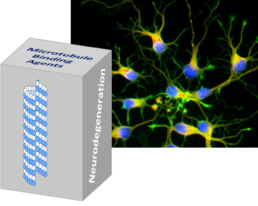
Microtubule-targeting agents and neurodegeneration
The association of microtubule (MT) breakdown with neurodegeneration and neurotoxicity has provided an emerging therapeutic approach for neurodegenerative diseases. Tubulin binders are able to modulate MT dynamics and, as a result, are of particular interest both as potential therapeutics and experimental tools used to validate this strategy. Here, we provide a comprehensive overview of current knowledge and recent advancements regarding MT-targeting approaches for neurodegeneration and evaluate the potential application of MT-targeting agents (MTAs) based on available preclinical and clinical data.
https://www.sciencedirect.com/science/article/pii/S135964462030516X
Tubulin tails and their modifications regulate protein diffusion on microtubules
Microtubules (MTs) are essential components of the eukaryotic cytoskeleton that serve as “highways” for intracellular trafficking. In addition to the well-known active transport of cargo by motor proteins, many MT-binding proteins seem to adopt diffusional motility as a transportation mechanism. However, because of the limited spatial resolution of current experimental techniques, the detailed mechanism of protein diffusion has not been elucidated. In particular, the precise role of tubulin tails and tail modifications in the diffusion process is unclear. Here, using coarsegrained molecular dynamics simulations validated against atomistic simulations, we explore the molecular mechanism of protein diffusion along MTs. We found that electrostatic interactions play a central role in protein diffusion; the disordered tubulin tails enhance affinity but slow down diffusion, and diffusion occurs in discrete steps. While diffusion along wild-type MT is performed in steps of dimeric tubulin, the removal of the tails results in a step of monomeric tubulin. We found that the energy barrier for diffusion is larger when diffusion on MTs is mediated primarily by the MT tails rather than the MT body. In addition, globular proteins (EB1 and PRC1) diffuse more slowly than an intrinsically disordered protein (Tau) on MTs. Finally, we found that polyglutamylation and polyglycylation of tubulin tails lead to slower protein diffusion along MTs, although polyglycylation leads to faster diffusion across MT protofilaments. Taken together, our results explain experimentally observed data and shed light on the roles played by disordered tubulin tails and tail modifications in the molecular mechanism of protein diffusion along MTs.
https://www.tubintrain.eu/wp-content/uploads/2020/11/Levy.pdf
Two Antagonistic Microtubule Targeting Drugs Act Synergistically to Kill Cancer Cells
Paclitaxel is a microtubule stabilizing agent and a successful drug for cancer chemotherapy inducing, however, adverse effects. To reduce the effective dose of paclitaxel, we searched for pharmaceutics which could potentiate its therapeutic effect. We screened a chemical library and selected Carba1, a carbazole, which exerts synergistic cytotoxic effects on tumor cells grown in vitro, when co-administrated with a low dose of paclitaxel. Carba1 targets the colchicine binding-site of tubulin and is a microtubule-destabilizing agent. Catastrophe induction by Carba1 promotes paclitaxel binding to microtubule ends, providing a mechanistic explanation of the observed synergy. The synergistic effect of Carba1 with paclitaxel on tumor cell viability was also observed in vivo in xenografted mice. Thus, a new mechanism favoring paclitaxel binding to dynamic microtubules can be transposed to in vivo mouse cancer treatments, paving the way for new therapeutic strategies combining low doses of microtubule targeting agents with opposite mechanisms of action.
https://www.tubintrain.eu/wp-content/uploads/2020/10/2020-Perrone.pdf
Structural Basis of Noscapine Activation for Tubulin Binding
Noscapine is a natural alkaloid that is used as an antitussive medicine. However, it also acts as a weak anticancer agent in certain in vivo models through a mechanism that is largely unknown. Here, we performed structural studies and show that the cytotoxic agent 7AO-demethoxy-amino-noscapine (7A-aminonoscapine) binds to the colchicine site of tubulin. We suggest that the 7A-methoxy group of noscapine prevents binding to tubulin due to a steric
clash of the compound with the T5-loop of α-tubulin. We further propose that the anticancer activity of noscapine arises from a bioactive metabolite that binds to the colchicine site of tubulin to induce mitotic arrest through a microtubule cytoskeleton-based mechanism.
https://www.tubintrain.eu/wp-content/uploads/2020/10/2020-Oliva.pdf
N‑alkylisatin‑based microtubule destabilizers bind to the colchicine site on tubulin and retain efficacy in drug resistant acute lymphoblastic leukemia cell lines with less in vitro neurotoxicity
Background: Drug resistance and chemotherapy-induced peripheral neuropathy continue to be significant problems in the successful treatment of acute lymphoblastic leukemia (ALL). 5,7-Dibromo-N-alkylisatins, a class of potent microtubule destabilizers, are a promising alternative to traditionally used antimitotics with previous demonstrated efficacy against solid tumours in vivo and ability to overcome P-glycoprotein (P-gp) mediated drug resistance in
lymphoma and sarcoma cell lines in vitro. In this study, three di-brominated N-alkylisatins were assessed for their ability to retain potency in vincristine (VCR) and 2-methoxyestradiol (2ME2) resistant ALL cell lines. For the first time, in vitro neurotoxicity was also investigated in order to establish their suitability as candidate drugs for future use in ALL treatment.
Methods: Vincristine resistant (CEM-VCR R) and 2-methoxyestradiol resistant (CEM/2ME2-28.8R) ALL cell lines were used to investigate the ability of N-alkylisatins to overcome chemoresistance. Interaction of N-alkylisatins with tubulin at the the colchicine-binding site was studied by competitive assay using the fluorescent colchicine analogue MTC. Human neuroblastoma SH-SY5Y cells differentiated into a morphological and functional dopaminergic-like neurotransmitter phenotype were used for neurotoxicity and neurofunctional assays. Two-way ANOVA followed by a Tukey’s post hoc test or a two-tailed paired t test was used to determine statistical significance.
Results: CEM-VCR R and CEM/2ME2-28.8R cells displayed resistance indices of > 100 to VCR and 2-ME2, respectively. CEM-VCR R cells additionally displayed a multi-drug resistant phenotype with significant cross resistance to vinblastine, 2ME2, colchicine and paclitaxel consistent with P-gp overexpression. Despite differences in resistance mechanisms observed between the two cell lines, the N-alkylisatins displayed bioequivalent dose-dependent cytotoxicity to that of the parental control cell line. The N-alkylisatins proved to be significantly less neurotoxic towards differentiated SH-SY5Y cells than VCR and vinblastine, evidenced by increased neurite length and number of neurite branch points. Neuronal cells treated with 5,7-dibromo-N-(p-hydroxymethylbenzyl)isatin showed significantly higher voltage-gated.
https://www.tubintrain.eu/wp-content/uploads/2020/10/Keenan.png
Structural model for differential cap maturation at growing microtubule ends
Microtubules (MTs) are hollow cylinders made of tubulin, a GTPase responsible for essential functions during cell growth and division, and thus, key target for anti-tumor drugs. In MTs, GTP hydrolysis triggers structural changes in the lattice, which are responsible for interaction with regulatory factors. The stabilizing GTP-cap is a hallmark of MTs and the mechanism of the chemical-structural link between the GTP hydrolysis site and the MT lattice is a matter of debate.
We have analyzed the structure of tubulin and MTs assembled in the presence of fluoride salts that mimic the GTP-bound and GDP.Pi transition states. Our results challenge current models because tubulin does not change axial length upon GTP hydrolysis. Moreover, analysis of the structure of MTs assembled in the presence of several nucleotide analogues and of taxol allows us to propose that previously described lattice expansion could be a post-hydrolysis stage involved in Pi release.
https://www.tubintrain.eu/wp-content/uploads/2020/10/Estevez-Gallego.png
On the Importance of the Thiazole Nitrogen in Epothilones: Semisynthesis and Microtubule- Binding Affinity of Deaza-Epothilone C
Deaza-epothilone C, which incorporates a thiophene moiety in place of the thiazole heterocycle in the natural epothilone side chain, has been prepared by semisynthesis from epothilone A, in order to assess the contribution of the thiazole nitrogen to microtubule binding. The synthesis was based on the esterification of a known epothilone A-derived carboxylic acid fragment and a fully synthetic alcohol building block incorporating the modified side chain segment and subsequent ring-closure by ring-closing olefin metathesis. The latter proceeded with unfavorable selectivity and in low yield. Distinct diferences in chemical behavior were unveiled between the thiophene-derived advanced intermediates and what has been reported for the corresponding thiazole-based congeners. Compared to natural epothilone C, the free energy of binding of deaza-epothilone C to microtubules was reduced by ca. 1 kcal/mol or less, thus indicating a distinct but non-decisive role of the thiazole nitrogen in the interaction of epothilones with the tubulin/microtubule system. In contrast to natural epothilone C, deaza-epothilone C was devoid of antiproliferative activity in vitro up to a concentration of 10 uM, presumably due to an insufficient stability in the cell culture medium.
https://www.tubintrain.eu/wp-content/uploads/2020/10/2020-Edenharter.pdf

